
Seasons come and go. Life is a journey with many detours. The big story of my life has many chapters in it yet to be written. Just when I thought I had climbed one summit and sat down to rest, a voice inside tells me, “Stand up, pick up your mat and go.… Read more
Category: ENT & Surgery
How to treat a nose bleed
Nose bleeds (epistaxis) is extremely common. Not every patient requires cauterisation. About 97% of all nose bleeds occur at the front of the nose on the septum (midline) as there are major vessels that arise from the floor of the nose to supply the septum.… Read more
Decisions before birth
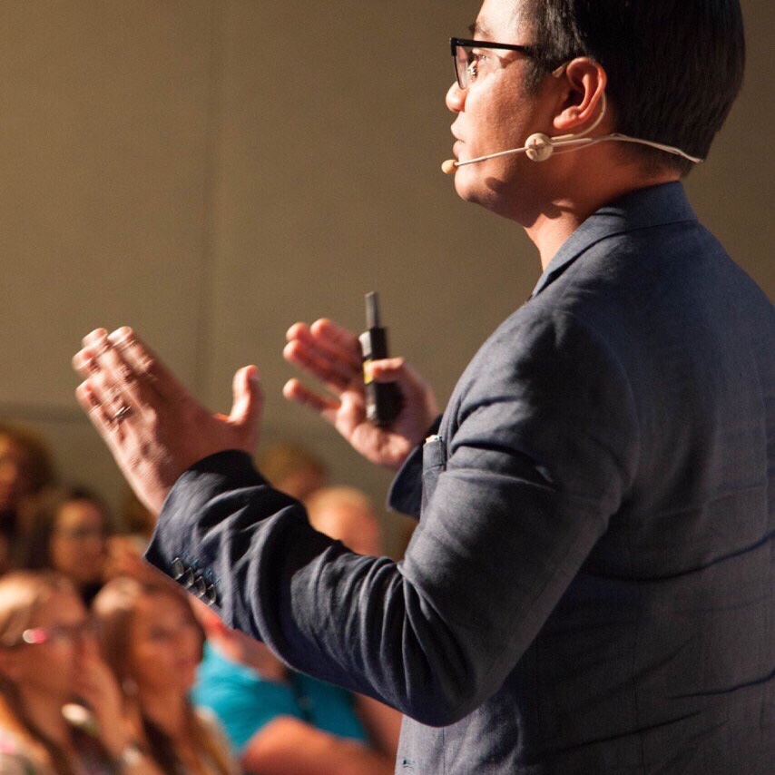
This talk was presented on the 13th of September at #CODA22.
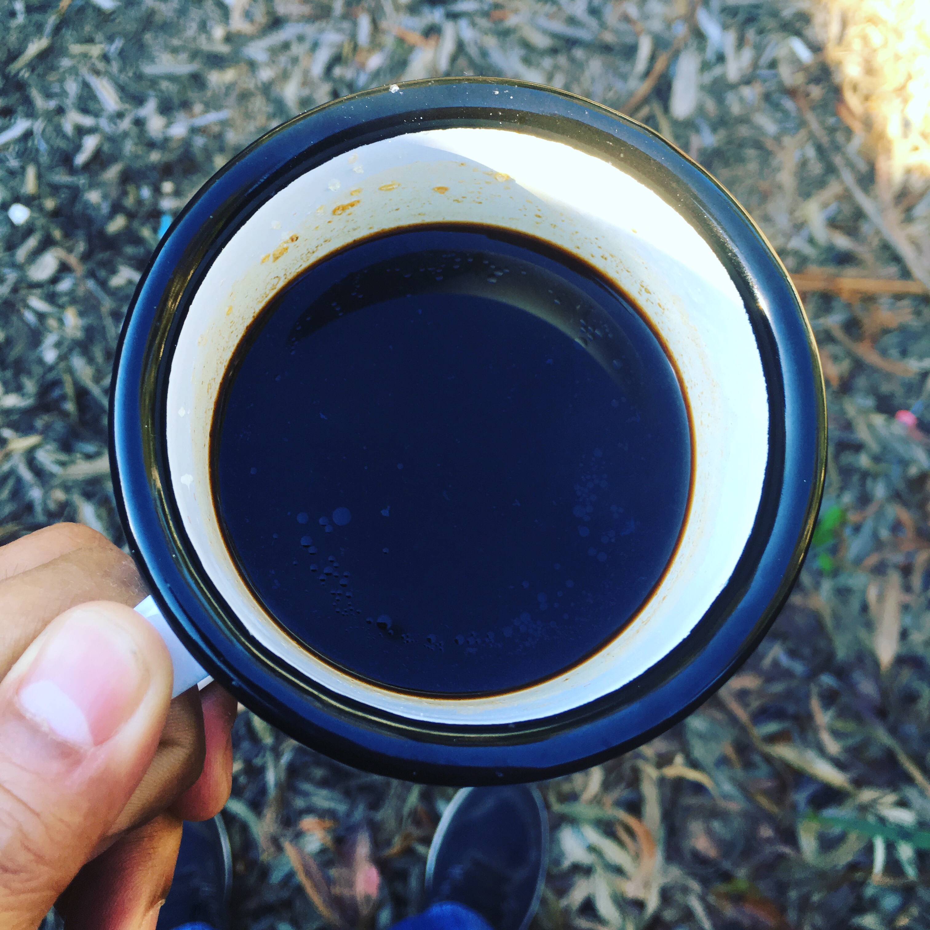
Wominjeka. Welcome. We make hundreds of decisions every single day. What you wear, who you speak to, latte, piccolo, magic, soy, almond, oat milk, lactose free, or if you’re not from Melbourne, black or white coffee.… Read more
Loss of Smell and Taste with COVID19
 Let’s get some terminology correct. COVID19 is the disease. SARS-CoV2 is the specific name given to the actual new virus that has been identified as the cause of COVID19. SARS-Cov2 is a new novel virus from the Coronavirus family. In lay terms, coronavirus is the general term we use to refer to this virus.… Read more
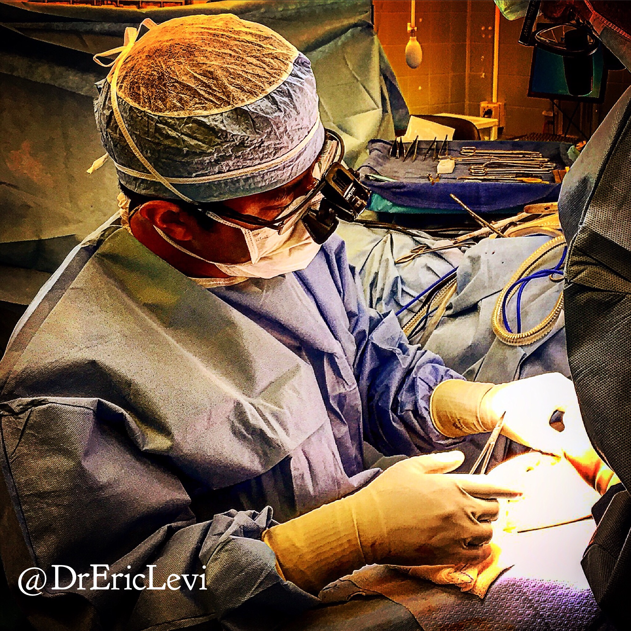
Nasopharyngeal swab and nasogastric insertion.
The nasopharynx goes back, not up.
When you take a nasopharyngeal swab or insert nasogastric tube, NEVER aim upwards towards the brain.
Go LOW and go SLOW.
Correct direction follows the floor of your nose not up towards the roof.… Read more
Loss of smell and taste with COVID19
 Here are a few fancy ENT words for you:
Olfactory (smell) disorder:
Anosmia: No smell (I can’t smell coffee)
Hyposmia: Reduced smell (I can smell coffee faintly)
Parosmia: Smelling a different smell (This coffee smells different)
Phantosmia: Smelling something that isn’t there (There’s a coffee smell but no coffee)
Gustatory (Taste) disorder:
Dysgeusia: dysfunction of taste
Parageusia: distortion of taste
Hypogeusia: reduced taste
Ageusia: No taste
ALSO, did you know that humans have 5 kinds of taste: sweet, salty, bitter, sour, umami (taste of MSG)
Read here from Europe the latest and so far the biggest study on smell and taste disorder in association with COVID19.… Read more
Airway ENT Talk at DFTB17
A presentation I gave at Don’t Forget The Bubble 2017 Acute Paediatric Conference On Brisbane, Australia.
Watch this if:
1. You’ve ever wondered how I actually sound in real life (don’t laugh)
2. You’re interested in the paediatric #ENTsurgery #Airway smorgasboard
Prof Graeme Clark & The Bionic Ear

It was an honour to interview the man who dedicated his life to pushing the boundaries of medical innovation. Prof Clark is the inventor of the cochlear implant, a medical marvel affectionately also known as the Bionic Ear. The idea that you could give hearing to the deaf is astounding.… Read more
World Head and Neck Cancer Day 2017
 July 27th is World Head & Neck Cancer Day.
What is a Head & Neck Cancer? Head & Neck Cancer is a diverse group of cancers that can arise from anywhere above the collar bones and outside the brain. They can occur from the skin, glands, soft tissue or the aerodigestive tract of the head & neck.… Read more
ORL2017 Surgeons and Social Media
ORL2017 Surgeons and Social Media
Presentation given at ORL2017 Honolulu, Hawaii, USA.… Read more